The Visual Cortex
Brief review of the visual cortex and the visual maps in the cortex.
Human visual system
During visual perception, light entering the eye is detected by the retina, an array of photoreceptors and related cells on the inside of the rear surface of the eye. The cells in the retina encode the light levels at a given location as patterns of electrical activity in neurons called ganglion cells. Output from the ganglion cells travels through neural connections to the lateral geniculate nucleus of the thalamus (LGN). From the LGN, the signals continue to the primary visual cortex (V1). V1 is the first cortical site of visual processing; the previous areas are termed subcortical. The output from V1 goes on to many different higher cortical areas, including areas that underlie object and face processing.
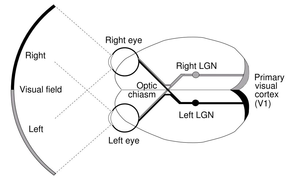
Human visual pathways (top view). Diagram of the main feedforward pathways in the human visual system.
Lateral Geniculate Nucleus (LGN)
The Lateral Geniculate Nucleus of the thalamus is the first step of visual processing. The LGN’s neurons do a process akin to edge detection between bright and dark areas. Some neurons at the LGN prefer bright over dark, or vice versa, being called ON or OFF cells.

ON cell in retina or LGN

OFF cell in retina or LGN
Primary Visual Cortex (V1)
The V1 is the first cortical site of visual processing. Input from the thalamus goes through afferent connections to V1, and the output goes on to many different higher cortical areas, including areas that underlie object and face processing. The neurons form local connections within V1 (long-range lateral connections) or connect to higher visual processing areas.
The cells themselves can prefer several features, for example, preferring one eye or the other, the orientation of the stimulus and its direction of movement, color combinations(such as red/green or blue/yellow borders), disparity (relative positions on the two retinas), etc.

Receptive field in V1. Starting in V1, most cells in primates have orientation-selective RFs. The V1 RFs can be classified into a few basic spatial types. The figure shows a two-lobe arrangement, favoring a \(45^\circ\) edge with dark in the upper left and light in the lower right
At a given location on the cortical sheet, the neurons in a vertical section through the cortex respond most strongly to the same eye of origin, stimulus orientation, spatial frequency, and direction of movement. It is customary to refer to such a section as a column . Nearby columns generally have similar, but not identical, preferences; slightly more distant columns have more dissimilar preferences. Preferences repeat at regular intervals (approximately 1–2 mm) in every direction, which ensures that each type of preference is represented across the retina. This arrangement of preferences forms a smoothly varying map for each dimension.
Visual maps
The term visual map refers to the existence of a non-random relationship between the positions of neurons in the visual centers of the brain (e.g. in the visual cortex) and the values of one or more of the receptive field properties of those neurons . The term is usually qualified by reference to the property concerned. For example, stimulus orientation is represented across the cortex in an orientation map of the retinal input. In an orientation map, each location on the retina is mapped to a region on the map, with each possible orientation at that retinal location represented by different but nearby orientation-selective cells. The figure below displays an example orientation map from monkey cortex.
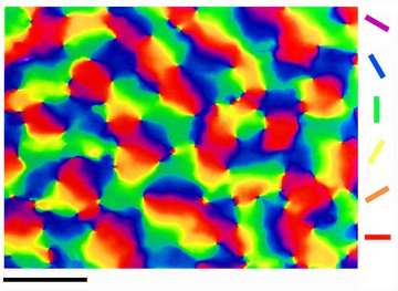
Orientation map in the macaque. Orientation preference map observed in area V1 of macaque monkey. Each neuron is colored according to the orientation it prefers, using the color key on the right. Nearby neurons in the map generally prefer similar orientations, forming groups of the same color called iso-orientation patches. Scale bar = 1 mm.